
J. R. Maxfield, MD
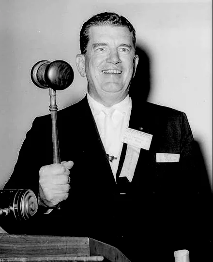
Eleventh president of the Southwestern Chapter (term ending 1967)
Dr. J.R. Maxfield grew up in a medical family. His father had begun the use of x-ray in 1903 and continued to practice radiology through the years. J.R. went to Baylor University in Waco, Texas where his father had moved to continue his medical practice by taking over the medical practice of his grandfather, George Dallas Streeter, M.D. and to continue in the field of radiology.
At the end of the second year of college at Baylor University in Waco, J.R. transferred to Baylor University College of Medicine, which was then in Dallas, Texas. After his second year in medical school at Baylor in Dallas, he was forced to drop out temporarily as this was the height of the depression. J.R. was employed as a Texas Ranger for approximately two years and obtained enough money to return to medical school. He continued all of his life to be an honorary Texas Ranger, a title of which he was very proud.
Before returning to medical school, J.R. and his brother Jack (who served as the second president of the Southwestern Chapter) went to Los Angeles, California. While in Hollywood, they met Howard Hughes and other notables. From Hollywood, they went to Chicago, Illinois where they became managers of the Texas exhibit at the Chicago World’s Fair in 1933. The story is told that on their way to Chicago they stopped for a nap in lower Illinois. When they awoke, their car was surrounded by Illinois State Police. It seems there had been a robbery, and the getaway car was similar to their car. When the Illinois State Troopers spotted their car, they were surprised to find both occupants wearing six-guns (see picture). J.R. showed them his Texas Ranger badge and instead of being arrested they were challenged to a shooting match with the best marksman on the Illinois State Police force. When J.R. and Jack won the shooting match, as a prize they were put in touch with the operators of the Chicago World’s Fair and landed the job as managers of the Texas exhibit.
Upon completing his medical school training at Baylor in 1935, Dr. J.R. interned at Port Chester Hospital in Port Chester, New York, 1935 to 1936, before returning to Baylor University Hospital to take his residency in radiology. As a radiology resident at Baylor, 1936 to 1938, Dr. J.R. worked with Charles Martin, M.D. where they developed the concept of the low intensity radium needles. Use of the low intensity needles significantly improved the response to radiation implant therapy of malignancy. Previously the high intensity needles that had been used, frequently caused severe radiation necrosis in the area implanted.
After completing his residency in 1938, Dr. J.R. took a fellowship in radiology at the University of California in San Francisco (UCSF). The radiology department at UCSF was unique because it had the first mega voltage radiation therapy unit, a one million volt x-ray therapy unit. While in San Francisco, Dr. J.R. became acquainted with the Lawrence brothers, Ernest Lawrence, PhD and John Lawrence, M.D. Ernest Lawrence was the physicist who had invented the cyclotron. He had installed a large cyclotron at Berkeley. It was John Lawrence, M.D., who in the late 1930s, started the field of clinical nuclear medicine using the radioactive isotopes I-131 and P-32 produced in his brother, Ernest’s, cyclotron. Dr. John Lawrence was an internist, but he worked closely with the radiology department at the University of San Francisco in California; therefore, my brother, J.R., had the opportunity of working with radioactive phosphorus P-32 and radioactive I-131 in 1938-39.
Upon completing this fellowship at the University of California in 1939, Dr. J.R., accepted a position as radiologist for the expanding Lovelace Clinic in Albuquerque, New Mexico. He worked there as a general radiologist for a year before returning to Dallas, Texas in 1941 to accept a position in radiology at Parkland Hospital. He maintained his contacts with the Lawrence brothers and their physicist friends who operated other cyclotrons. Therefore, he was able to obtain radioactive iodine and radioactive phosphorus during the World War II years, enabling him to start clinical nuclear medicine with nuclear medicine treatment and diagnostic programs at Parkland through the department of radiology. During World War II, Dr. J.R. was also a consultant to Los Alamos National Laboratory where he had the opportunity to meet Edward Teller, PhD and many of the other individuals involved in the Manhattan Project, which is a separate story in itself.
Drs. J.R. and Jack Maxfield established the Maxfield Clinical-Hospital at 2711 Oak Lawn, Dallas, Texas, in 1947, incorporating a nuclear medicine laboratory into the hospital as one of the first Texas hospitals to have a functioning nuclear medicine laboratory. They also had one of the first Atomic Energy Commission approvals for a nuclear medicine program. Their brother, William Maxfield, was privileged during the summer, on weekends, and during odd times to work as a nuclear medicine technician in that facility. At that time, they had only Geiger-Mueller (GM) counters, and for high level monitoring, the QTPIE unit. The cutie pie monitor was developed for the Manhattan Project. The story told is that they were looking for a code name for the monitor. Part of the formula for the detection chamber was QT π, hence the name of QTPIE.
Scintillation crystal system detectors were not developed until the early 1950's. At the Maxfield Clinic-Hospital, they were using radioactive iodine-131 for diagnosis of thyroid problems and for treatment of hyperthyroidism and thyroid cancer. One of the interesting cancer patients was a young child with pulmonary metastasis from an undifferentiated thyroid cancer. The uptake of her undifferentiated thyroid cancer of I-131, as expected, was very low. Therefore, she was treated with an unusual program of 1 MCI of I-131/day, 5 days per week for 300 doses. She had an eight-year survival. The Maxfield Clinic-Hospital also used radioactive phosphorus P-32 for treatment of leukemia, and other blood dyscrasias. We also developed the technique of using P-32 as a diagnostic procedure similar to the sentinel node biopsies of today. Prior to surgical resection of the colon, colon cancer patients would be given 2 MCI of radioactive phosphorus (P-32) intravenously. After resection of the colon cancers, they would do an autoradiograph of the surgical specimen, which would be obtained before sending the surgical specimen to the pathologist. This technique could find hot spots of P-32 localization on the autoradiograph. As a result, they could tell the pathologist where to go to make pathologic sections to confirm the presence of metastatic disease if the initial pathology report was negative for lymph node metastasis. This study with autoradiograph P-32 technique showed that in about 30% of the cases, we could demonstrate metastatic nodes in patients who had been given a clean bill of health for metastasis on the original routine pathology report. Unfortunately, it does not seem that this data was ever published. Another unusual use of P-32 was treatment of brain cancer. P-32 with a needle type GM counter had been used at surgery to assist in removal of brain tumors by checking the margin of the tumor for radioactivity. We used P-32 to treat brain cancers that had failed standard therapy. One example is an eight year-old boy who had a brain stem medulloblastoma that had not responded to external radiation therapy. With a total of 20 MCI of P-32 given 2 MCI/day, 5 days a week we achieved a 13-year survival for this patient with failed radiation therapy.
Dr. J.R. and Dr. Jack recognized the need for a low cost nuclear medicine scanner that was also space efficient. With their physicist friend, they developed the prototype 3” scintillation crystal scanner that was manufactured and sold by Picker as the Clini Scanner. This unit had a number of firsts. It was the first unit to use a preset window for the various isotopes. Also the unit had a teledeltos paper readout system. The unit measured only about 3’ x 3’ for floor area. While an excellent clinical scanner, the Clini Scanner never achieved wide utilization as at the time it was introduced the trend in nuclear medicine was to the camera system which could provide function studies as well as static scans.
At the Maxfield Clinic-Hospital, they installed a Tracer Lab ½ inch scintillation crystal nuclear scanner and in August of 1955 installed in their facility the third Cobalt 60 Radiation Therapy unit in the U.S. for radiation therapy treatment. Having worked with the 1 million volt x-ray therapy unit at the University of California, Dr. J.R. avoided the pitfalls of super voltage radiation therapy encountered by many radiologists when they converted to tele cobalt 60 or linear accelerator radiation therapy.
After the Society of Nuclear Medicine and the Southwestern Chapter of Nuclear Medicine had been in operation for a number of years, it was recognized that there was a need for another spokesman for the field of nuclear medicine which would be predominantly physician based. This need for a physician based spokesman for the field occurred when the Airline Pilots Association threatened to stop shipment of radioactive materials on passenger carrying planes after an accident when a technetium generator leaked causing contamination of the plane. Dr. J.R. asked his friend and patient, Edward Teller, PhD, to go to Washington, D.C. to talk to the Airline Pilots Association, the senators and congressmen about the importance of nuclear medicine and need to continue distribution of radiopharmaceuticals on passenger carrying aircraft. This incident with the generator leak occurred before the arrival of Fed-Ex and other commercial freight carriers to serve smaller communities. It was comments from these meetings that preserved use of passenger carrying aircraft to distribute radiopharmaceuticals that there which highlighted the need for a physicians based group to speak for the field of nuclear medicine. The Maxfields, therefore, became in 1971 the incorporators of the American College of Nuclear Medicine, which has provided a sound basis of clinically oriented scientific programs for practitioners of the field of nuclear medicine through the years.
Some additional notes from an August 1997 “In Memoria” article published in the Journal of Nuclear Medicine by Thomas P. Haynie, MD:
- Martin Nusynowitz, MD remembered Dr. J.R. at the annual business meeting of the Southwestern Chapter in New Orleans on April 12, 1997 as a man of "presence" and the quintessential Texan who dressed the part with boots, a wide brimmed hat and a diamond stickpin. He was a Charter Member and the Secretary-Treasurer of the Southwestern Chapter for many years and its 11th president. His brother Jack was the chapter's second president, and his brother William, now of Florida, was the chapter's 17th president.
- John Hidalgo, MS, SNM past president from New Orleans, remembers Dr. Maxfield's noteworthy contributions to the SNM. He hired the first administrator of the SNM, Sam Turiel of Chicago, and it was during his presidency that The Journal of Nuclear Medicine got started, evolving from a quarterly to a bimonthly and then a monthly, doubling the number of pages. He instituted the Nuclear Pioneer Lecture and invited Dr. Edward Teller to give the lecture at the 1960 SNM meeting in Estes Park, CO. He hosted SNM meetings in Dallas in 1962 and again in 1976.
- Dr. J.R. was an avid supporter of the Space Program, he served on many NASA committees and attended rocket launches from Friendship 7 to the Space Shuttle.
- Dr. J.R.'s passing recalls the exciting early years of the field of nuclear medicine. Here was a man who grasped the importance of the field at an early stage, studied with its pioneers and gave freely of his time and energy to help direct its growth during its critical years of development.